Extras din referat
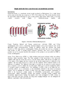
Dividing cells undergo a series of shape changes that begin in mitosis and continue throughout cytokinesis. In this experiment, we used AFM to study shape changing during cell division. Atomic force microscopy (AFM) can be performed simultaneously with optical microscopy techniques, such as fluorescence or differential interference contrast (DIC) microscopy. Compared to other method, we can study the living cells under physiological conditions without fixation and chemical staining. More important, we can also measure the force produced by the dividing cells based on the force-distance equation: F= k•x. The force produced in this process is in the range of N.
Experimental procedure
Cell culture
1. Incubate HeLa cells with GFP on cover slips; the culturing medium was DMEM
2. Wash HeLa cells grown on cover slips several times with 500μL PBS
3. Select the suitable cover slip using optical microscopy. The following parameters should be considered:
a. Density of cells per cover slip →___ not too high, otherwise it wouldn’t be possible to pick up and analyze only one cell
b. Density of cells undergoing mitosis per cover slip →___ high enough so that it wouldn’t be too difficult to find a proper subject in the sample
4. The selected cover slip was transferred in a small dish with DMEM
AFM
1. Position the laser on the cantilever
2. Transfer the cover slip in the sample chamber
3. Calibrate the system (touch the surface with the cantilever for 10s with nm force) to reduce the thermo fluctuation
a. this step is necessary to discriminate between force caused by shape changing or heat drift
4. Find a cell in prophase by exciting the GFP labeled cells with 508nm laser light
5. Touch the selected cell with the cantilever that has the following features:
a. no tip is present because it could destroy cell membrane upon changing its shape
b. a short cantilever (the shorter the stiffer) is selected because the length of the cantilever may influence the force-distance equation
Discussion
During mitosis the changes upon genetic material and upon cell itself are well known and documented. The AFM experiment was a very concise verification of this standard knowledge. The DIC images allowed the observation of how the shape of the cell changed during the process, assuming a virtually perfect round shape before metaphase and consequently splitting at the end of the M phase.
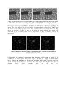
Fluorescence microscopy permitted the observation of DNA during cell division by labeling the histone H2A. As expected, the loosely bundled chromatin initially condensed and shaped itself into chromosomes (individual chromosomes were not distinguished, of course). This is the stage known as Prophase. Needless to say, by this point the genetic material has already been duplicated. At this point the nuclear envelope disassembles to allow microtubules to reach the kinetochores.
In Metaphase, the condensed chromosomes align themselves roughly along the middle of the rounded cell, as shown by the fluorescence images in the form of a bright, broad vertical line. This is followed in Anaphase by chromosome separation and motion in opposite directions, rapidly followed by Telophase and cytokinesis. The DIC images show the formation of a tiny cleavage furrow which eventually splits the cell into two.
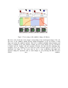
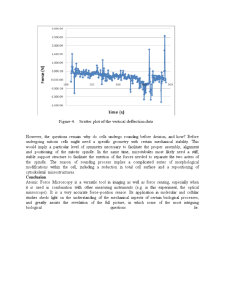
Figure 3: Force acting on the cantilever during cell division.
The force curve should also show changes corresponding to the morphological change of the cell. The force on the cantilever rapidly increases during prometaphase, as the cell acquires a round shape and pushes the cantilever upwards. This force is stabilized gradually during metaphase and reaches a maximum in the early anaphase. Further, the force drops quickly once the cell division is finished and the daughter cells get separated. However, the data (text file mentioning time, elongation, force) we obtained does not match the results observed during the execution of the experiment (see Figure 4). The reason may be due to some modifications that might have appeared during the experiment such as small changes in cell positioning will affect the force exerted upon the cantilever
Preview document


Conținut arhivă zip
- Investigation of Cell Globularization During Mitosis by Combining Optical Microscopy and Atomic Force Microscopy.doc